
Reading Time: mins
Last Updated: 09/14/26
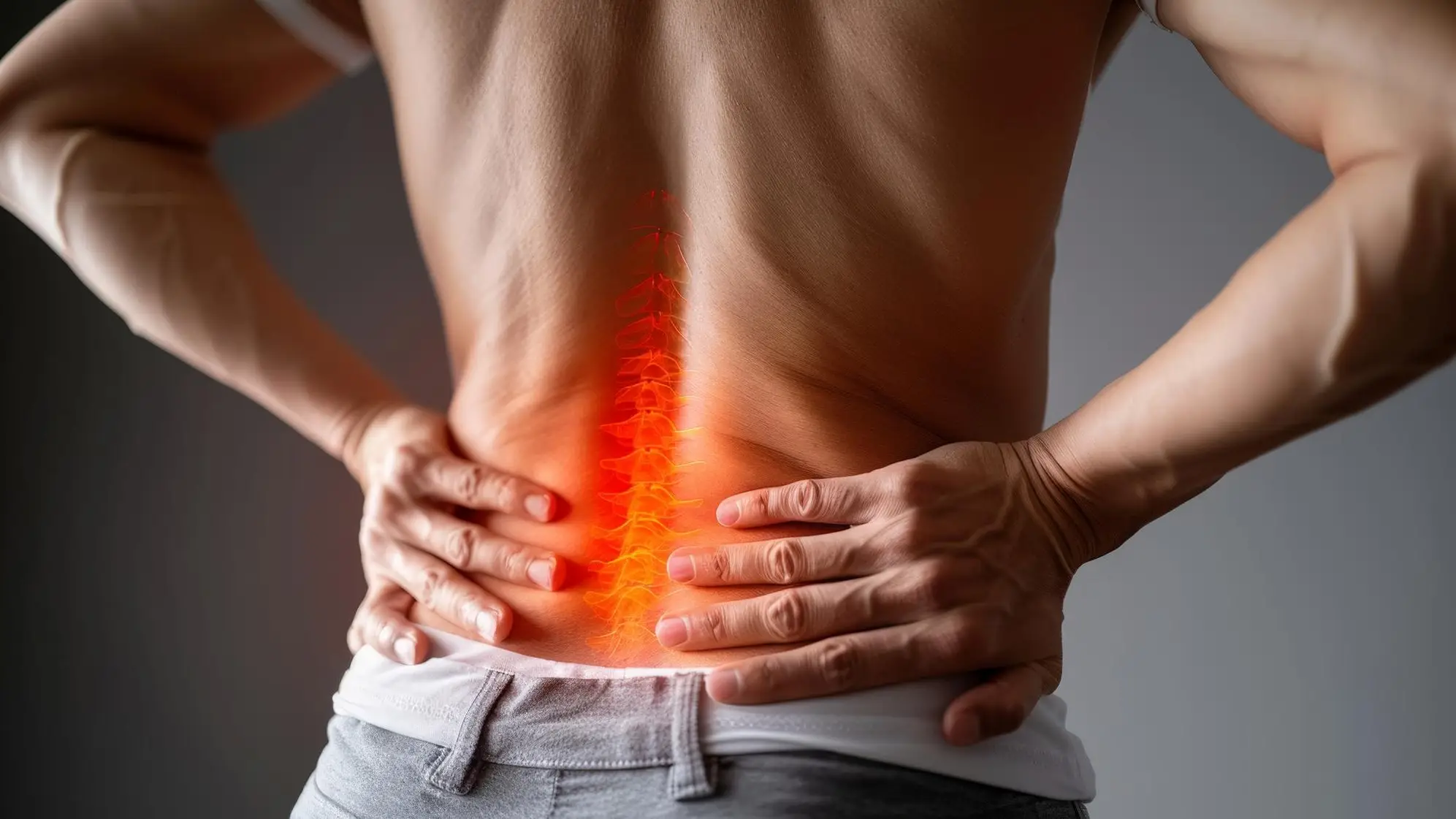
If you have been told you have disc degeneration in the lower back and are picturing an operating table, take a breath first. Most cases never need surgery at all. At RRMG, we see this diagnosis often, and in the vast majority of patients we can manage the pain, protect movement, and keep people active using injections, targeted rehab, and lifestyle changes alone. This guide walks through what the finding means, why it happens, what your scan grade tells you, and the non-surgical path we use to treat it in London, plus answers to the questions we hear most in clinics.
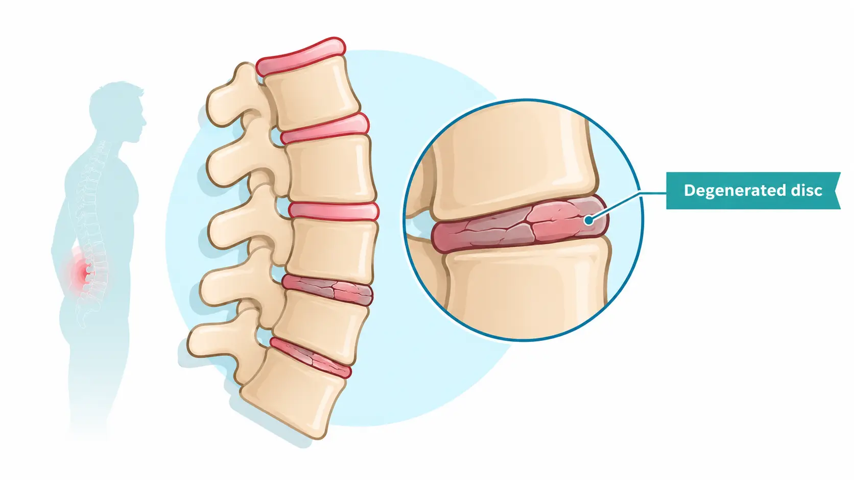
Intervertebral disc degeneration is the gradual breakdown of the cushioning discs that sit between the bones of your spine. Each disc has a tough outer ring, the annulus fibrosus, wrapped around a soft, gel-like centre called the nucleus pulposus. Together the two parts absorb shock and let your spine bend and twist without the bones grinding against each other.
Over time, that gel centre loses water, the outer ring can develop small tears, and the disc loses some of its height and cushioning ability. This is disc degeneration meaning in plain terms: your discs ageing and drying out, much like a shock absorber wearing down after years of use. It is a slow, mechanical process rather than an infection or a sudden injury, which is part of why it can develop quietly for years before anyone notices symptoms.
Let’s be plain about this: disc degeneration is not the same thing as a slipped or herniated disc, and it is not automatically a disease. A 2015 review of MRI scans in people with no back pain at all found disc changes in a large share of adults over 40, rising further with age. Structural change on a scan does not always mean pain, and pain does not always match how a scan looks. If you have just received this diagnosis, that mismatch should offer real reassurance: a scan finding is one piece of information, not a verdict. We explain what your specific scan means for you and your daily life, rather than reading the report in isolation, and we take the time to answer the questions a printed report never covers, such as why you feel pain on some days and not others.
Doctors commonly grade disc degeneration on a five-point scale first described by radiologist Christian Pfirrmann in 2001, based on how a disc appears on an MRI scan. It remains the standard reference point today, and it is the same scale we use when reviewing your imaging with you.
Types of disc degeneration are usually described by where the breakdown shows up rather than by a separate disease name. Some people mainly lose disc height. Others develop small tears in the outer ring, bony spurs at the disc edges (a pattern sometimes called lumbar spondylosis), or narrowing of the space the disc normally fills. Several of these changes often appear together in the same person, which is one reason two people with the same grade on a scan can feel very different day to day.
This matters clinically because grade alone does not decide your treatment plan. A Grade IV disc in someone with good core strength and no nerve involvement can be far less troublesome than a Grade II disc sitting next to an irritated nerve root. We treat the person and the pattern of symptoms in front of us, using the grade as context rather than as the whole answer. That is also why two patients handed the same MRI report can walk away with two very different treatment plans, and why we would rather spend extra time on assessment than fit every scan into a single template.
Disc degeneration in the lower back is largely a normal part of aging, but a handful of factors speed it up or make it more likely to cause pain. The main contributors we see in clinic are:
Types of disc degeneration are usually described by where the breakdown shows up rather than by a separate disease name. Some people mainly lose disc height. Others develop small tears in the outer ring, bony spurs at the disc edges (a pattern sometimes called lumbar spondylosis), or narrowing of the space the disc normally fills. Several of these changes often appear together in the same person, which is one reason two people with the same grade on a scan can feel very different day to day.
This matters clinically because grade alone does not decide your treatment plan. A Grade IV disc in someone with good core strength and no nerve involvement can be far less troublesome than a Grade II disc sitting next to an irritated nerve root. We treat the person and the pattern of symptoms in front of us, using the grade as context rather than as the whole answer. That is also why two patients handed the same MRI report can walk away with two very different treatment plans, and why we would rather spend extra time on assessment than fit every scan into a single template.
Disc degeneration in lower back is largely a normal part of ageing, but a handful of factors speed it up or make it more likely to cause pain. The main contributors we see in clinic are:
Disc degeneration age is a fair question, because timing varies more than people assume. Early changes can start showing up on scans from the late twenties onward, and men tend to show measurable disc changes around a decade earlier than women, although women can go on to experience more pronounced effects later in life, particularly after menopause when hormonal changes affect disc tissue directly. By midlife, some degree of disc change on imaging is common rather than unusual, which is exactly why we weigh a scan alongside your actual symptoms rather than treating the image as the whole story.
Disc desiccation is the specific term for the water loss inside a disc’s centre, and it is one of the earliest signs a disc is degenerating. On an MRI, a well-hydrated disc shows up bright on certain scan sequences. As the disc dries out, that brightness fades to grey and eventually to a dark, dehydrated appearance, which is usually the first change a radiologist will note on your report.
Desiccation on its own does not always cause pain. It is a description of what the disc looks like, not a diagnosis of a painful condition, and many people with desiccated discs on a scan have no symptoms whatsoever. Where it does contribute to pain, it is usually because the drier, flatter disc is less able to absorb load evenly, which can irritate nearby joints and soft tissue over time and change how weight is shared between spinal segments during everyday movement.
This is one of the most common findings we see mentioned on an MRI report handed to a worried patient, often with no further explanation attached. Radiologists note desiccation because it is visible and easy to describe, not because it automatically signals a problem that needs treating. Part of our first consultation is simply walking through the report line by line, so you leave understanding which findings are routine and which ones are driving your symptoms.
Disc desiccation lumbar changes specifically affect the discs in your lower spine, the region that carries the most day-to-day load from standing, walking, bending, and lifting. Because the lumbar discs work harder than discs higher up the spine, desiccation here is common and often shows up earlier than in the neck or mid-back. When we see lumbar desiccation on a scan alongside axial low back pain, movement stiffness, or pain that eases with rest, we look at it as one piece of a bigger picture rather than the single explanation, since axial low back pain often involves several overlapping structures working together, including the facet joints and the deep stabilising muscles either side of the spine.

Disc degeneration treatment at RRMG almost always starts with a proper assessment: a review of your scan, a physical examination, and a conversation about what limits your day, before any treatment is suggested. From there, we build a plan around the least invasive option that fits your specific presentation, and move up only if that is not enough. This step-by-step approach matters because two people with an identical MRI finding can need very different plans depending on their fitness, work demands, and how their pain behaves day to day.
Our confirmed non-surgical toolkit for lower back disc degeneration includes:
| Approach | What it targets | Typical relief window |
| Steroid injection | Inflammation around the disc or joint | Short to mid-term |
| Medial branch block | Confirms the pain source (diagnostic) | Not intended for lasting relief |
| Radiofrequency ablation | Facet or SI joint-mediated pain | Around 6 to 12 months or more |
| Transforaminal epidural steroid injection | Nerve root swelling from sciatica | Short to mid-term |
| PRP | Supports the disc’s own healing response over time | Builds over several sessions |
None of these treatments regrow a disc back to how it looked at twenty, and we will always tell you that plainly rather than overselling a result. What they can do is calm pain, restore movement, and help you avoid or delay surgery for a condition that, for most people, responds well to a conservative approach. We built this diagnose-first approach into how we treat every case, confirming a pain source with a medial branch block before moving to radiofrequency ablation, drawing on founder Dr Ralph Rogers‘s years of image-guided musculoskeletal work at the top level of professional sport. That sequencing, diagnose first and only then treat, is deliberate: it stops a patient going through a procedure aimed at the wrong joint or the wrong level, which is a common reason non-surgical care fails elsewhere before it even gets a fair chance.
Ultrasound and fluoroscopy-guided steroid injections settle inflammation around the disc or a nearby joint quickly, often within a few days of treatment. We use live imaging for every injection so the medication lands on the exact structure driving your symptoms rather than spreading through the surrounding tissue. Relief typically runs from a few weeks to a few months, which is usually enough of a window for rehab to build lasting strength and control around the affected level. We treat this as a bridge into that rehab work rather than a repeatable fix on its own, since leaning on injections alone tends to mask the underlying mechanical problem instead of addressing it.
A medial branch block is a diagnostic step before it is a treatment. We inject a small amount of local anaesthetic around the nerves supplying a specific facet joint, then track whether that block removes your pain, even briefly. If it does, we have confirmed that joint as a real source of your symptoms and can move forward with radiofrequency ablation at that same level with actual evidence behind the decision, rather than treating a joint we have only guessed is involved.
Radiofrequency ablation uses heat delivered through a fine needle-tip electrode to disable the small nerve supplying a facet or sacroiliac joint, carried out only once a medial branch block has confirmed that joint as the pain source. Relief commonly lasts six to twelve months or more, since the treated nerve takes time to regrow and start signalling pain again. It is a day-case procedure done under local anaesthetic with light sedation, and most patients return to normal daily activity within a day or two.
Transforaminal epidural steroid injections place anti-inflammatory medication directly around an irritated nerve root, the structure usually behind sciatica-type leg pain when disc degeneration is involved. We guide the needle to the exact spinal level under live imaging rather than injecting blind. Relief tends to sit in the short to mid-term range, long enough to calm an acute flare while rehab and movement retraining deal with the underlying mechanical load that triggered it.
Disc degeneration pain relief usually combines more than one approach rather than relying on a single fix. A short course of anti-inflammatory medication or a guided steroid injection can settle an acute flare, while ongoing relief tends to come from rebuilding strength and control around the spine so the affected level carries less strain day to day. Heat, gentle movement, and pacing your activity through a flare also help more than strict bed rest, which can leave the lower back stiffer once the initial pain settles. Most patients notice the biggest shift not from any single treatment, but from the combination of a calmer flare and steadier, more consistent movement habits afterwards.
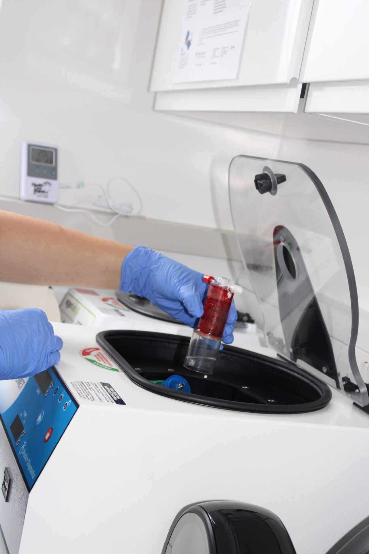
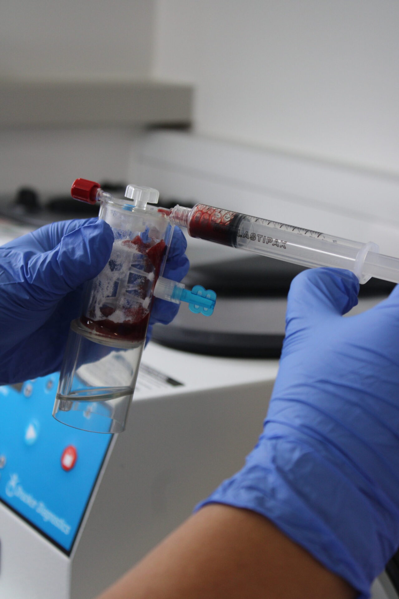
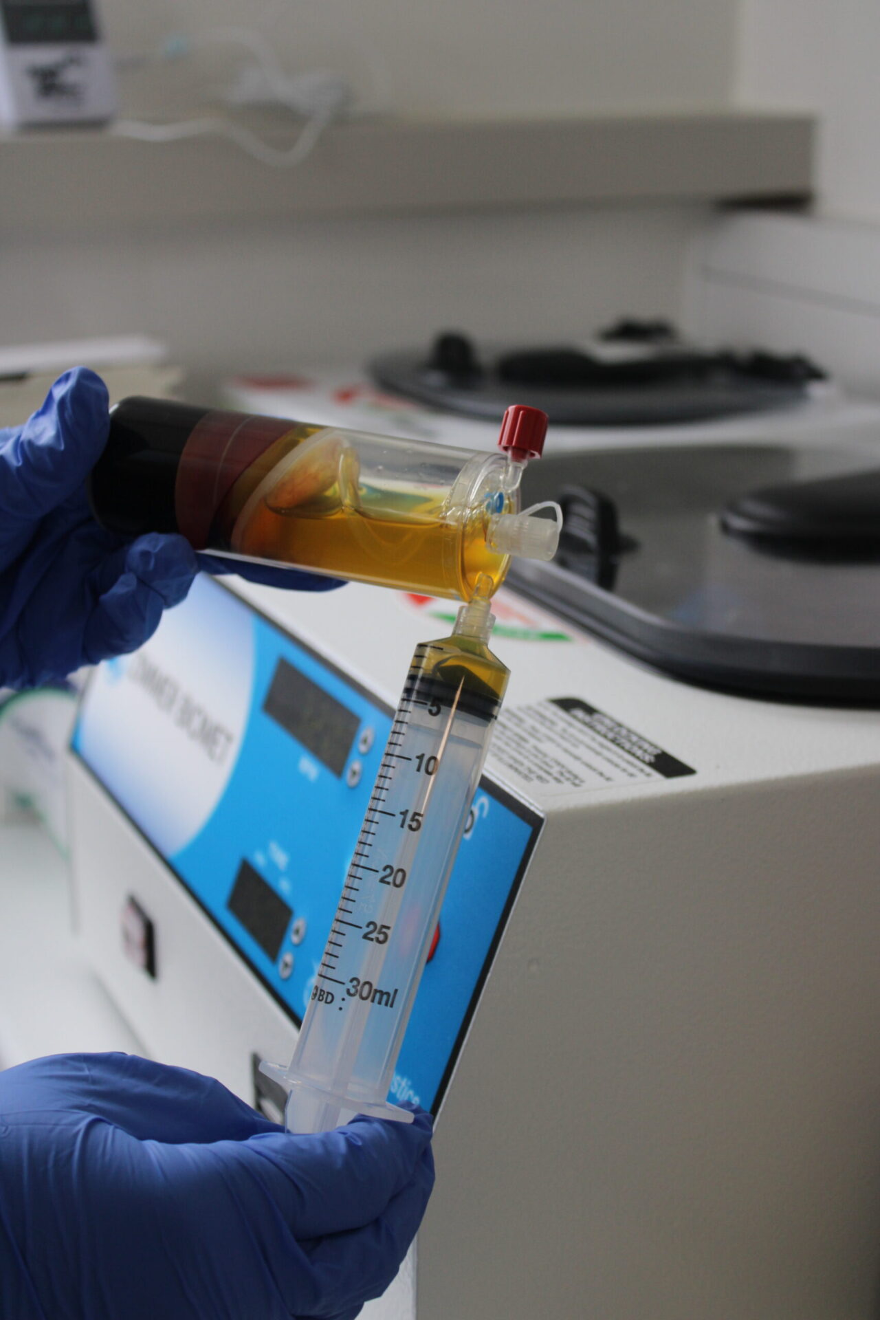
PRP for disc degeneration uses a concentrated sample of your own blood platelets, injected near the affected area to support the body’s natural repair response. It will not rebuild a disc from scratch, and we are careful never to promise that. For the right patient, particularly someone looking for a longer-term, regenerative-leaning option alongside rehab rather than repeated steroid injections, it is a legitimate part of the non-surgical toolkit worth discussing at your consultation. We will talk you through whether your specific pattern of degeneration is one where PRP tends to add real value, rather than offering it as a default add-on to everyone who asks.

We are RRMG, a private regenerative orthopaedics and sports medicine clinic on Blythe Road in London, founded by Dr Ralph Rogers, whose case we personally run from your first scan review through to your treatment plan. We work with a wide range of insurance partners, as well as welcoming self-funding patients, so cost should never be the reason you put off getting a proper assessment.
If disc degeneration in lower back is affecting your daily life, work, or sport, we would like to help you understand exactly what your scan means and which of the options above is the right fit for your case. Our lower back spinal injections page has more detail on each treatment, and our fees page has current guide pricing for a first consultation.
People often ask us this alongside a related question, can you fix disc degeneration once it shows up on a scan. The honest answer to both is no: disc degeneration cannot be fully stopped or fixed once it has started, since it reflects the natural ageing of the disc itself. What can change is its pace and its impact: staying active, keeping a healthy weight, not smoking, and treating flares early with the right non-surgical care can slow further wear and keep symptoms well controlled for years, often long enough that the underlying grade on a scan matters far less than how well your spine actually functions day to day. Patients who stay consistent with rehab and pacing tend to report fewer and shorter flares over time, even when their scan grade has not changed at all.
Disc desiccation on its own is rarely serious. It is a normal, common finding on MRI scans, especially from midlife onward, and plenty of people with desiccated discs have no pain at all. It becomes more clinically relevant only if it is paired with red flag symptoms such as bladder or bowel changes, saddle numbness, or unexplained weight loss, which need urgent medical assessment rather than routine back pain care.
Disc space narrowing happens when a disc loses height as it dries out and its structure changes with wear. Ageing is the biggest driver, alongside genetics, long-term mechanical load from posture and lifting habits, smoking, and carrying extra body weight, all of which can speed up how quickly a disc’s height reduces over time.
Disc space narrowing is usually not serious by itself and is a common feature of an ageing spine rather than a diagnosis that demands urgent action. It becomes more important when it is severe enough to reduce the space around a nerve root, which can bring on leg pain, numbness, or weakness that should be assessed properly rather than left to settle on its own.
Disc space narrowing is treated the same way as broader disc degeneration: rehab and movement retraining first, with guided injections, medial branch blocks, or radiofrequency ablation added when a specific pain source is confirmed and needs more targeted relief. Surgery is reserved for the small number of cases where nerve compression is severe and conservative care has already been given a fair chance to work.
For more on how RRMG approaches spine pain without surgery, see our related post on non-surgical spine solutions, or read the NICE guidance on low back pain that shapes our conservative-first approach.
Dr Rogers has had a rich and illustrious career so far, which is continuing to grow and develop. Everything Ralph has been involved in since qualifying has added value to his knowledge and experience, but there have been some impressive highlights to note:
2018 – Present Founder Medical Director: Rogers Regenerative Medical Group (RRMG)
2012 – 2024 Medical Advisor – National Basketball Association
2008 – 2018 Medical Director: The London Sports Injury Clinic London
2010-2011 First Team Doctor, Chelsea Football Club
2007- 2010 The Football Association (Team Physician for England under 19 and development squads)
2005-2006 Medical Advisor: Trinidad & Tobago World Cup Football Team
2002 – 2008 Club Doctor: Walsall Football Club, West Midlands England
1998 – 2009 Medical Director: Medical & Orthopaedic Sports Therapy,
2000 – 2005 Sports Physician: Warwickshire County Cricket Club, Birmingham