
Reading Time: mins
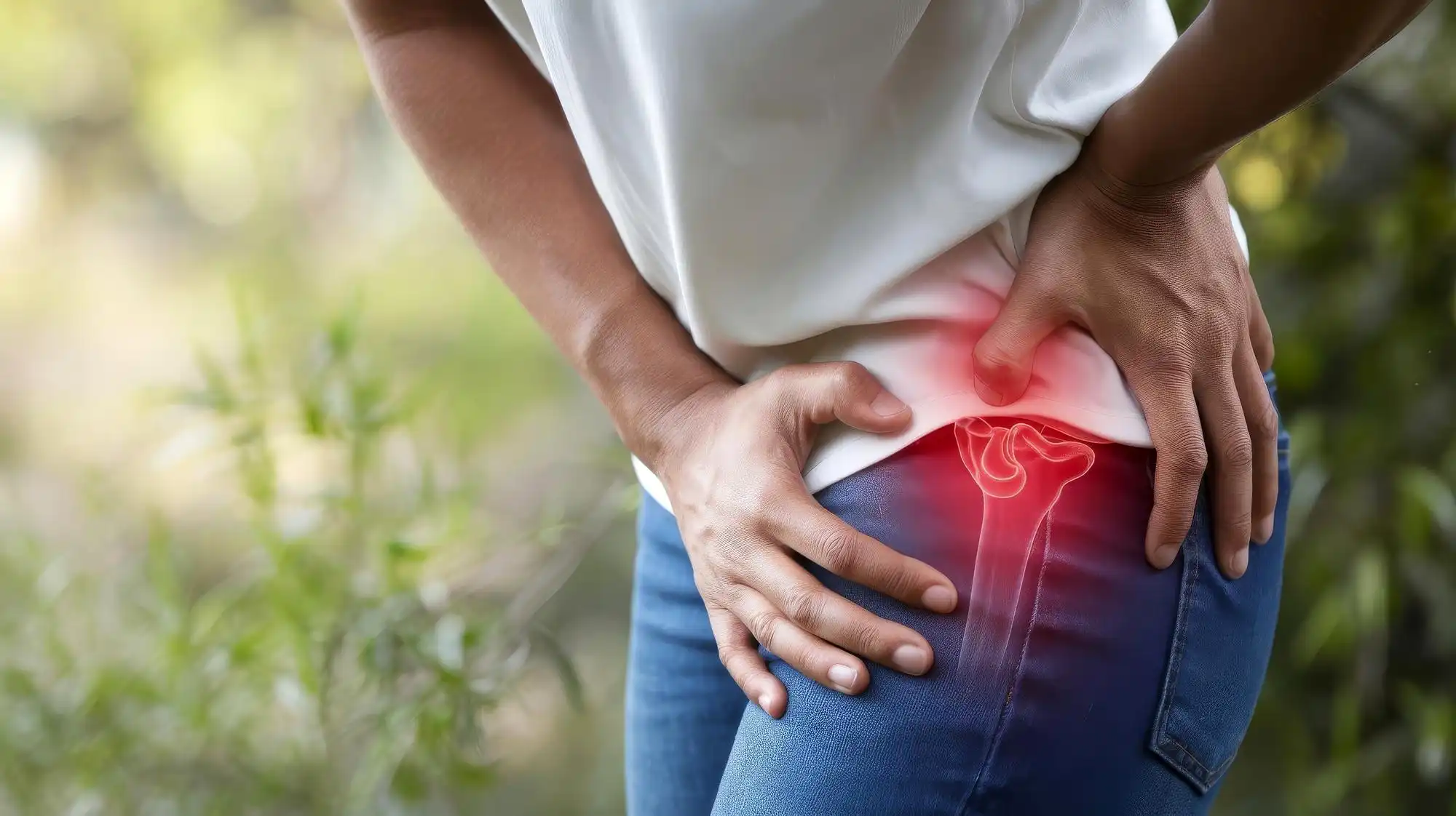
If you’ve been told you have hip bursitis, you may have noticed the advice you’re given doesn’t always match what a male friend or relative was told for the “same” condition. That’s not inconsistency on your doctor’s part. Female hip bursitis treatment genuinely looks different in practice, because the condition itself behaves differently in women, from who it affects to which structures are actually involved. At Rogers Regenerative Medical Group, our image-guided approach is built around that distinction rather than a one-size-fits-all injection.
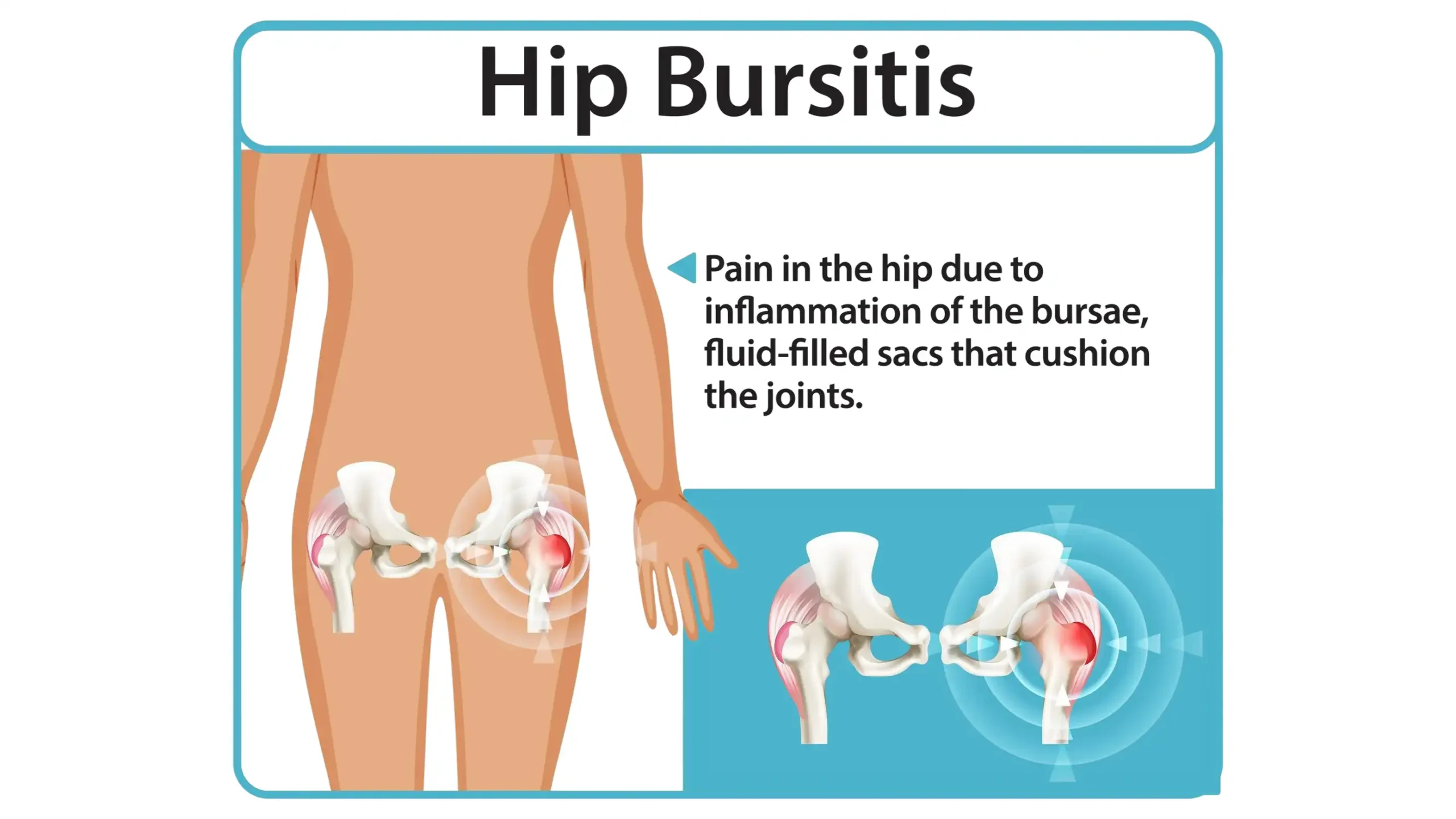
Bursitis happens when a bursa, one of the small, fluid-filled sacs that sit between bone, muscle, and tendon to reduce friction, becomes inflamed. You have bursae throughout your body wherever soft tissue glides over bone, and the hip has several. Hip bursitis specifically involves the bursa that sits over the greater trochanter, the bony point on the outer side of your hip where several tendons and the iliotibial band pass closely over the bone. When that bursa is irritated, everyday movements like walking, climbing stairs, or lying on your side can become noticeably uncomfortable, sometimes seemingly out of proportion to how minor the triggering activity felt at the time.
The condition can develop gradually, from repeated low-level friction building up over weeks or months, or it can follow a single specific incident, such as a fall directly onto the hip. Either way, once the bursa is inflamed, it tends to stay sensitive until the underlying irritation, whether that’s a mechanical issue, an activity pattern, or tendon involvement, is properly addressed rather than just calmed down temporarily.
We increasingly describe this as part of a wider picture called greater trochanteric pain syndrome, rather than hip bursitis inflammation in isolation. That’s an important shift: it means the bursa is rarely the whole story, and it’s a large part of why treatment for hip bursitis has moved beyond a single steroid shot and nothing else. Understanding that distinction is also the first step toward understanding why the condition, and its treatment, so often looks different in women.
The core symptoms of hip bursitis are the same regardless of sex, but women are considerably more likely to experience them in the first place, and often present with a broader symptom pattern than the bursa alone would explain.
The defining symptom is pain directly over the point of the hip, on the outer thigh. Most people describe it as a dull, achy pain rather than a sharp one, though it can flare with certain movements. It’s common to notice:
Some women notice what feels like a distinct lump or firm swelling over the outer hip rather than a general ache. In most cases this is the inflamed bursa itself, or thickened, irritated tendon tissue sitting close to it, rather than anything more serious. It can feel alarming to find a new lump, especially if it appeared suddenly or seems to shift slightly with movement.
We should be upfront that this specific “lump” description is our own plain-language read on how bursal swelling tends to present, not a phrase drawn from a clinical source the way the rest of this section is. Any new lump around a joint is still worth having examined properly rather than assumed. Imaging is the only reliable way to tell bursal swelling apart from a tendon tear, a cyst, or another cause, and self-diagnosing from symptoms alone is one of the more common ways treatment ends up delayed or mismatched to what’s actually happening underneath.
Understanding hip bursitis causes is the fastest way to work out why your own symptoms keep coming back. A number of everyday and structural factors can trigger the condition, and most of them are things you can actually do something about:
Once the bursa is irritated, certain positions and activities reliably make it worse: lying on the affected side, sitting with your legs crossed for long periods, climbing stairs repeatedly, and any high-impact activity like running on hard surfaces. Even simple daily habits, such as always sleeping on the same side or sitting on a low, soft sofa that puts the hip into a compressed position for long stretches, can quietly keep a flare going without an obvious trigger.
Carrying extra body weight also increases the mechanical load on the hip and is associated with a meaningfully higher risk of developing the condition in the first place, which is one reason activity and weight management are usually part of a treatment plan rather than an afterthought. None of this means these activities need to be avoided permanently, but recognising which of them line up with your own flares is often the fastest way to reduce how often symptoms recur.
Diagnosis starts with a clinical examination: we press around the point of your hip to check exactly where the tenderness is, and ask about what movements and positions bring symptoms on. For most people, that clinical picture is enough to start treatment.
Imaging is used to rule out other causes and to see what’s actually going on beneath the surface. Plain X-rays help rule out hip arthritis or bone spurs, both of which can cause similar-feeling hip pain but need a different treatment plan entirely. Ultrasound lets us see the bursa and surrounding tendons in real time, which is especially useful for guiding treatment precisely to the affected structure rather than injecting by feel. MRI gives the most detailed picture of all three and is the most reliable way to tell simple bursal inflammation apart from gluteal tendon damage. That distinction matters: the two can need meaningfully different treatment approaches, and getting it right the first time can save months on a plan that was only ever addressing half the problem.
This is the part that genuinely sets female hip bursitis symptoms and treatment apart, and it comes down to hard prevalence data, not assumption. Research on greater trochanteric pain syndrome shows it affects up to 15% of women, compared with roughly 8% of men, a two- to five-fold difference. Onset most often peaks between ages 40 and 60, and carrying additional body weight further raises the risk.
That prevalence gap matters clinically for two reasons. First, we should have hip bursitis and gluteal tendon involvement genuinely on the list when evaluating hip pain in a woman in this age range, not treated as an afterthought once more common explanations are ruled out. Second, modern imaging has shown that what gets labelled “hip bursitis” is very often gluteal tendon damage sitting alongside the bursal inflammation, not the bursa acting alone. Treatment that only addresses the bursa can miss that tendon component entirely. That’s a large part of why a course of injections alone doesn’t always hold for as long as patients expect.
Higher body weight adds a second, separate risk factor on top of the sex difference. The two can compound for some patients rather than sit independently. None of this is destiny, and it doesn’t mean every woman with hip pain will develop bursitis. It’s part of why a treatment plan built around “this is what usually works” tends to under-deliver compared with one built around “this is what’s actually going on in your hip.”
That combination of factors is why we treat this presentation as its own conversation rather than defaulting to the same single-injection approach for everyone. Picture a woman in her late 40s with months of lateral hip pain and a previous injection that only helped briefly. Now picture a younger male athlete with a recent-onset flare and no prior treatment. Both might get referred to a clinic with the same word, “bursitis,” on the letter, but they are statistically very different clinical pictures.
At RRMG, ultrasound-guided steroid injections are one of our core non-surgical treatments for bursitis, delivered directly to the inflamed area under real-time imaging rather than by feel. Patients typically notice improvement within days, with relief that can last weeks to months. We’re upfront about the trade-off: published research shows steroid injections for this condition are effective, but they also carry a meaningfully higher chance of the pain returning within five years compared with other approaches. That’s exactly why we don’t treat an injection as a standalone fix.
For patients dealing with the gluteal tendon component described above, we also use regenerative options such as PRP (platelet-rich plasma), designed to support the tendon’s own healing response rather than simply mask inflammation, and shockwave therapy, which uses focused sound wave pulses to stimulate the tissue. Combined with a structured exercise programme rather than used alone, these approaches have shown strong results in published research. We’ll always be clear about what’s evidence-based versus what’s still emerging, and we never promise a guaranteed outcome for any individual case. If you’re weighing PRP against other regenerative options, our PRP vs stem cell comparison covers the practical differences.
Conservative management, meaning activity changes, targeted physical therapy, and anti-inflammatory medication, resolves the large majority of cases without any injection at all, which is why we start most patients there before discussing procedures. In practice, that usually means a physiotherapist-led programme focused on strengthening the hip abductor muscles and gently stretching the IT band, since it’s the combination of a stronger, more resilient tendon and a less irritated bursa that tends to hold up over time, rather than calming the bursa down in isolation and hoping the underlying mechanics take care of themselves.
Where a patient has already tried rest, physiotherapy, and over-the-counter measures without lasting improvement, that’s usually the point where an image-guided injection or a regenerative option becomes worth discussing, rather than the first step. We’ll talk you through the realistic timeline and trade-offs for each option at your consultation, based on what your imaging actually shows, rather than moving straight to the same procedure for every presentation.
Alongside whatever we recommend at your consultation, a few simple habits genuinely help while the bursa settles down:
If home measures aren’t shifting things after a couple of weeks, or the pain is disrupting your sleep and daily activity, that’s the point to have it properly assessed rather than continuing to manage it alone. Pushing through pain for months on the assumption it will eventually settle on its own is one of the more common reasons a straightforward case of bursitis turns into a more complicated, longer-standing problem.

If hip pain has been dismissed as “just bursitis” without anyone checking whether the gluteal tendons are involved, or if a previous injection helped only briefly, it’s worth having it reassessed properly. That’s especially true if you recognise the pattern described above: pain that’s been present for months rather than weeks, a previous steroid injection that helped for a while and then wore off, or hip pain that started around your 40s or 50s without an obvious single injury behind it.
We bring over 25 years of experience in sports and regenerative medicine to every case, shaped by founder Dr Ralph Rogers’s years as an NBA medical advisor and Chelsea FC’s first-team doctor. We deliver every image-guided treatment personally, never handing it off to a junior colleague.
An initial consultation starts the process with a proper clinical and ultrasound assessment, so any treatment plan, whether that’s activity guidance, an image-guided injection, or a regenerative option like PRP, is based on what’s actually happening in your hip rather than a guess. That’s the same image-guided principle behind every treatment we offer: see the structure clearly first, then treat it precisely, rather than working from symptoms alone. Current starting prices for consultations and injections are listed on our fees page, and we work with a wide range of insurance providers as well as self-funding patients.
To arrange an assessment, request an appointment with our team, or call us directly on +44 (0)207 112 5400.
Yes, though the intensity varies. Most people describe a dull, persistent ache over the outer hip that sharpens with specific movements like climbing stairs or lying on that side, rather than a constant severe pain.
It depends on the approach. Conservative care such as activity changes and physical therapy typically needs a few months to fully settle symptoms. A steroid injection can bring noticeable relief within days, though as above, that relief doesn’t always hold long-term on its own.
It can, especially after a steroid injection used alone. That’s one reason we build a broader plan around addressing the underlying cause, including any gluteal tendon involvement, rather than relying on repeat injections.
The underlying condition is the same, but it’s substantially more common in women, roughly two to five times more likely, and more often involves the gluteal tendons alongside the bursa. That’s the main reason treatment approaches are increasingly tailored by presentation rather than applied identically to everyone.
No, though the two can feel similar and sometimes coexist. Hip bursitis is inflammation of a soft-tissue sac on the outer hip, while hip arthritis involves wear of the joint itself and tends to produce pain deeper in the groin as well as at the side. Imaging is what reliably tells the two apart, since the treatment for each is different; our separate guide on hip arthritis treatment covers that condition specifically.
Rogers Regenerative Medical Group is a private regenerative orthopaedics and sports medicine clinic based at Blythe Road, London, founded by Dr Ralph Rogers. This article is for general information and does not replace individualised medical advice.
Dr Rogers has had a rich and illustrious career so far, which is continuing to grow and develop. Everything Ralph has been involved in since qualifying has added value to his knowledge and experience, but there have been some impressive highlights to note:
2018 – Present Founder Medical Director: Rogers Regenerative Medical Group (RRMG)
2012 – 2024 Medical Advisor – National Basketball Association
2008 – 2018 Medical Director: The London Sports Injury Clinic London
2010-2011 First Team Doctor, Chelsea Football Club
2007- 2010 The Football Association (Team Physician for England under 19 and development squads)
2005-2006 Medical Advisor: Trinidad & Tobago World Cup Football Team
2002 – 2008 Club Doctor: Walsall Football Club, West Midlands England
1998 – 2009 Medical Director: Medical & Orthopaedic Sports Therapy,
2000 – 2005 Sports Physician: Warwickshire County Cricket Club, Birmingham